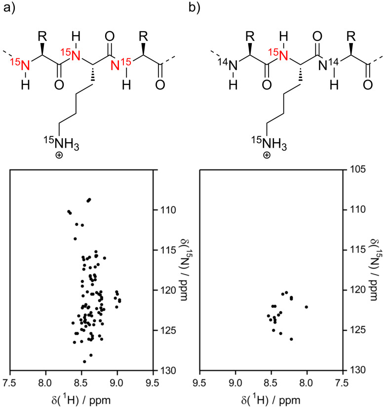
Figure 5.
Schematic 1H,15N-HSQC spectrum of tauF4 (chemical shifts from BMRB # 17945, [109]) with and without specific 15N-lysine labeling. (a) Uniformly 15N-labeled protein. The amide NH of all residues (except Pro) yield a signal, resulting in signal overlap. (b) Selective 15N-lysine labeling. Only the amide NH of lysine residues are visible. The specific labeling significantly reduces signal overlap and thus makes it easier to track the shifting of single resonances upon ligand binding.

