Figure 1.
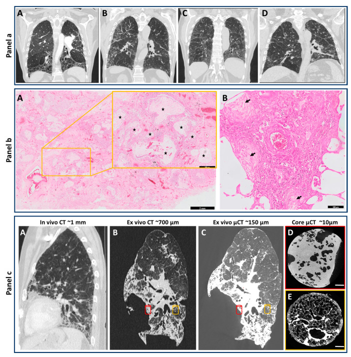
HRCT and Histopathological features for diagnosis of human UIP pattern, and overview of the potential use of CT and µCT for understanding the clinical pathology of end-stage IPF. Panel (a), HRCT: (A) Typical UIP in the setting of IPF. The presence of a peripheral, subpleural predominantly basal irregular reticular pattern, basal honeycombing and peripheral traction bronchiectasis makes IPF very likely; (B) Probable UIP in the setting of IPF. Peripheral and subpleural irregular reticular pattern with distal traction bronchiectasis but without honeycombing makes IPF very likely in case also clinical findings suggest this diagnosis; (C) Nonspecific interstitial pneumonia (NSIP). The dominant presence of ground-glass opacity together with a fine regular reticular pattern and proximal traction bronchiectasis makes IPF less likely; (D) Chronic fibrotic hypersensitivity pneumonitis. Irregular lines and reticular pattern are present throughout both lungs without peripheral dominance. In addition, lung density is inhomogeneous. IPF is unlikely and clinical arguments for an alternative diagnosis should be looked for. Panel (b): Histopathology of human lung (HE-staining). (A) Presence of honeycombing (inset, enlarged from yellow box, black stars) by formation of cystic airspaces of varying sizes filled with mucus and lined by bronchiolar epithelium (A scale bar = 2 mm, scale bar in inset = 500 µm); (B) Fibroblast foci showing interstitial ongoing fibrosis without inflammatory infiltrates are indicated by black arrows (scale bar = 100 µm). Panel (c): In vivo chest HRCT scan 6 months prior to transplantation with a resolution of around 1 mm (A); Ex vivo CT of the transplanted lung highlighting an increased spatial resolution due to the absence of breathing artefacts (B); Whole lung ex vivo µCT showing sagittal view with a resolution up to 150 µm (C); Core µCT with a resolution of 10 µm provides insight into different areas within the lower lobe showing severe fibrosis (red cylinder) and a near-normal area (orange cylinder) demonstrating the need for rigorous characterization of separate regions within the same lung specimen (D,E).

