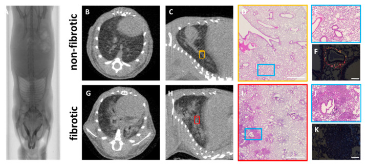
Figure 2.
Overview of the use of µCT and histology in a pre-clinical setting in a silica-induced lung fibrosis mouse model. Mice were oropharyngeally instilled with crystalline silica particles (5 mg/mouse, bottom row) or saline (top row). Whole-body µCT scan showing the lungs of a mouse instilled with silica to induce fibrosis (A). Axial (B,G) and sagittal (C,H) views of non-fibrotic and fibrotic lungs. Lung fibrosis is confirmed using histology at low magnification (12.5×—scale bar 2 mm) (D,I) and high magnification (50×–scale bar 0.5 mm—blue box) from areas indicated with yellow and red cylinders in (C,H), showing normal and dense fibrotic regions consisting of silica clustering, cellular infiltration and granuloma formation (E,J). Polarization microscopy (scale bar 0.1 mm) (F,K) allows visualization and quantification of the maturity of fibrotic lesions and silica clustering.

