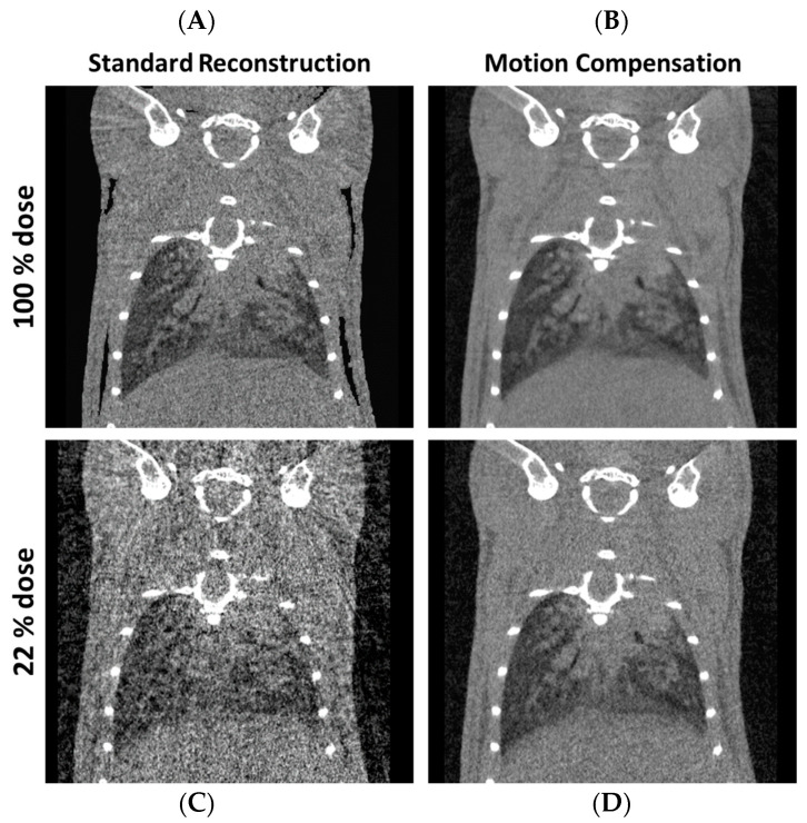
Figure 3.
Motion correction of in vivo µCT to improve image quality and dose exposure. Respiratory-gated 4D µCT reconstructions of a mouse with a respiratory window width of 20%. The top row shows reconstructions obtained with the full dose of the used reference protocol (A,B) and the bottom row shows reconstructions obtained using only 20% of the reference dose (C,D). The standard reconstruction (FBP) results in severe artifacts if dose is reduced while a motion compensation (MoCo) approach results in an image quality sufficient for most qualitative and quantitative tasks (D). (C = 50 HU, W = 400 HU). Videos are available as a supplementary: Video S1a and S1b.

