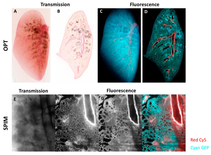
Figure 5.
Optical Projection Tomography (OPT) and Selective Plane Illumination Microscopy (SPIM) of silica-induced lung fibrosis. Mice were oropharyngeally instilled with crystalline silica particles (5 mg/mice), sacrificed and imaged 35 days after instillation. OPT transmission imaging of optically cleared lungs shown as a “raw” projection (A) and as a reconstructed slice (B). The regions with concentrations of silica particles can be seen as grey blotches in the transmission images (A,B). OPT fluorescent images of projected (C) and reconstructed (D) lungs. In fluorescence, the larger vascular structures can be seen in the Cy5 channel (red in images C,D), and the general structure is visualized using autofluorescence in the GFP channel (cyan in C,D). A more in-depth and spatial analysis of optically cleared lungs using SPIM shows a transmission image (E) and corresponding fluorescence optical slices in the Cy5 (F) and GFP (G) channels. (E) is a transmission image taken in the SPIM showing a field of view with regions containing silica particles (note that this is a traditional wide-field image, not an optical section). Optical sections of fluorescence are shown in (F–H), where regions with both normal structure and fibrosis can be seen. The images in (F,G) are shown in color overlay in (H). 3D visualizations of the OPT can be found as Supplementary Videos S3a and S3b.

