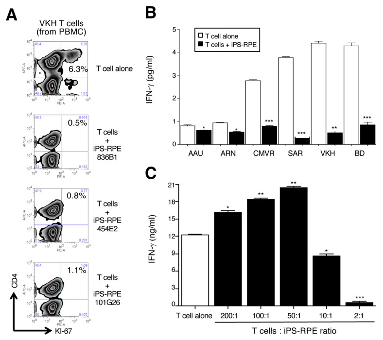
Figure 1.
Capacity of iPS-derived RPE to suppress or stimulate uveitis T cells in vitro. (A) Three lines of established human iPS-RPE cells were used (836B1, 454E2, and 101G26) for the in vitro assays. Uveitis T cells were established from PBMC of a VKH disease patient with active uveitis. The proliferation of CD4+ T cells in VKH PBMC in the presence of iPS-RPE cells was evaluated by FACS analysis (CD4 and Ki-67 staining). PBMC were cocultured with to 5 × 10⁵ (T cell:RPE ratio = 2:1) iPS-RPE cells. Numbers in the histograms indicate the percentage of cells double-positive for CD4 and Ki-67. (B) Data of IFN-γ-production in supernatants from Th1-type CD4+ uveitis T cells + iPS-RPE cells. T cells were cocultured with to 5 × 10⁵ (T cell:RPE ratio = 2:1) iPS-RPE cells. Open bars indicate T cells alone, and black bars indicate T cells plus iPS-RPE cells. AAU: HLA-B27 associated acute anterior uveitis; ARN: acute retinal necrosis; CMVR: CMV retinitis; SAR: sarcoidosis; VKH: Vogt-Koyanagi-Harada disease; BD - Behçet’s disease. Data are the means ± SEM of three ELISA determinations. (C) Capacity of iPS-RPE cells to suppress the activation of bystander T cells in a T-cell number assay. In the presence of antihuman CD3 agonistic antibody and rIL-2, purified CD4+ T cells from PBMC of active uveitis patients with VKH disease were cocultured with 5 × 10³ to 5 × 10⁵ (T cell:RPE ratio = 200:1, 100:1, 50:1, 10:1, or 2:1) iPS-RPE cells (454E2) for 48 h. Results indicate the IFN-γ production by T cells exposed to iPS-RPE cells. Data are the means SEM of 3 ELISA determinations. * p < 0.05, ** p < 0.005, *** p < 0.0005, as compared to the positive control (T cells alone: open bar).

