Figure 6.
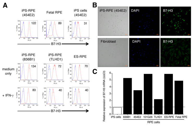
Detection of B7-H3 costimulatory molecules on human iPS-RPE cells. (A) The expression of B7-H3 on iPS-RPE cells was assessed by FACS analysis. 454E2 iPS-RPE cells, fetal RPE cells, and 454E2 iPS cells as a control were used. In the analysis, IFN-γ-pretreated RPE cells (iPS-RPE cells or ES-RPE cells) were also prepared. The numbers in the histograms indicate MFI. Blue histogram: data for isotype control. (B) Detection of B7-H3 on iPS-RPE cells by immunostaining. iPS-RPE cells (454E2) clearly expressed B7-H3 but fibroblasts did not. Cell nuclei were counterstained with DAPI. Scale bars, 50 μm. (C) In qRT-PCR for B7-H3 mRNA, PS-RPE cells, 836B1, 454E2, 101G26, TLHD1, ES-RPE cells, fetal RPE cells, and control iPS cells were prepared. Results indicate the relative expression of B7-H3 (ΔΔCt: control iPS cells = 1).

