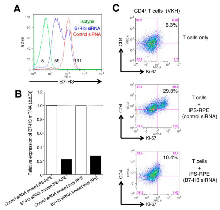
Figure 8.
Effect of B7-H3 expression by iPS-RPE cells on the stimulation of T-cell activation. (A) B7-H3-siRNA-transfected iPS-RPE cells (TLHD1) were harvested on day 3 and examined for expression of B7-H3 by flow cytometry. Control-siRNA-transfected cells were also analyzed. The numbers in the histograms indicate MFI. Data are representative of 2 experiments. (B) B7-H3-siRNA-transfected iPS-RPE cells (TLHD1) or control RPE cells (fetal RPE cells) were examined for expression of B7-H3 mRNA by qRT-PCR. Data are representative of 3 experiments. Control-siRNA-transfected cells were also analyzed (ΔΔCt: control cells = 1). (C) CD4+ T cells from a VKH disease patient were cocultured with B7-H3-siRNA-transfected iPS-RPE (or control-siRNA-transfected) cells and evaluated by Ki-67 staining (for proliferation) of T cells. T cells were cocultured with to 2 × 10⁴ (T cell:RPE ratio = 100:1) iPS-RPE cells. Data are representative of 3 individual experiments. Numbers in the histograms indicate the percentage of cells double-positive for CD4 and Ki-67.

