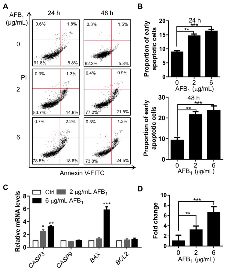
Figure 6.
AFB1 induces apoptosis in IMR-32 cells. (A) Apoptosis analysis of IMR-32 cells after AFB1 treatment by flow cytometry. The data were analyzed with FlowJo software. (B) The proportion of early apoptotic cells was calculated for the cells in panel A. (C) Fold changes in the mRNA levels of apoptosis-related genes in IMR-32 cells after exposure to 2 μg/mL or 6 μg/mL AFB1 for 24 h. All the mRNA levels tested were normalized to GAPDH. (D) The enzyme activity of caspase-3 in cells was detected via ELISA after treatment with 2 μg/mL or 6 μg/mL AFB1 for 24 h. All experiments were performed in triplicate and the values represent the mean ± SD of three independent experiments. Statistical significance was defined as * p < 0.05, ** p < 0.01, or *** p < 0.001.

