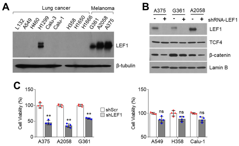
Figure 1.
Suppression of LEF1 attenuates melanoma cell growth. (A) LEF1 expression in lung cancer and melanoma cell lines. LEF1 protein levels were measured by using Western blotting. (B) LEF1 knock-down efficiency in melanoma cell lines. A375, A2058, and G361 melanoma cell lines were infected with lentivirus expressing shRNA against control or LEF1. The infected cells were selected by puromycin for six days. −, shRNA-scramble and +, shRNA-LEF1. (C) LEF1 knock-down attenuates melanoma cell growth but not lung cancer cells. LEF1 knocked-down melanoma and lung cancer cells were generated and cell viability was measured by crystal violet staining. The values represent the mean ± SD (n = 4); ns, not significant and ** p < 0.01. Red and blue points, mean value of individual sample.

