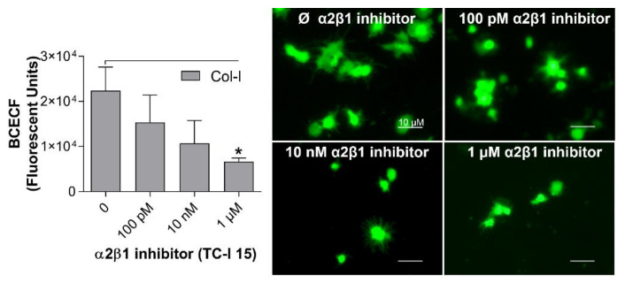
Figure 5.
Sensitivity of the optimized assay to detect platelet adhesion inhibition. Coating of 96-well microplates with collagen-I (4 µg/mL in 50 µL) or distillated water was performed for 1 h at 37 °C. After blocking the wells with BSA (0.03%), human washed platelets (8 × 10⁴/µL in 100µL) containing different concentrations of TC-I 15, an α2β1 integrin inhibitor, were added to the coated wells, followed by incubation for 1 h at 37 °C. Non-adherent platelets were removed, and adherent platelets were incubated with BCECF-AM (4 µg/mL in 50 µL) for 30 min at 37 °C. Fluorescence intensity was measured using a plate reader (VictorX, PerkinElmer). Values were compared to the control group without TC-I 15 by one-way ANOVA followed by Dunnett’s post hoc test (* p < 0.05, data are mean ± SEM; n = 4). Images were acquired with a fluorescence microscopy (Eclipse Ti2, Nikon, Waltham, MA, USA) with a 20x objective (scale bar 10 µM).

