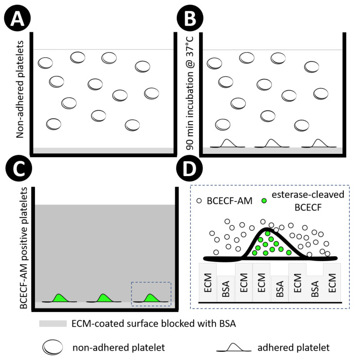
Figure 7.
Schematic representation of the study design using BCECF-AM. Extracellular matrix proteins were employed to coat 96- or 384-well microplates, followed by BSA blocking, both incubated for 1 h at 37 °C. Next, human washed platelets were added into wells (A). and incubated for 90 min at 37 °C (B). Non-adherent platelets were washed away with tyrode buffer, and adherent platelets were incubated with diluted BCECF-AM for 30 min at 37 °C. The excess of BCECF-AM was removed by washing the plate 3 times with tyrode buffer (C). Non-fluorescent BCECF-AM is permeable to the platelet membrane. Once inside the platelet, intracellular esterases cleave the ester bond, releasing BCECF, which is the fluorescent form of the molecule. In addition, the cleavage of lipophilic blocking groups by esterases, leads to a charged form of BCECF, which leaks out of cells more slowly than BCECF-AM [69]. Rectangular boxes represent ECM and BSA coating(D).

