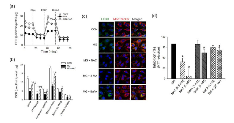
Figure 4.
Effects of NAC and autophagy inhibitors on mitochondrial damage induced by MG in brain ECs. (a,b) bEnd.3 cells were treated with NAC (5 mM) for 1 h before and during MG stimulation (1000 μM) for 6 h. Cells were applied to Seahorse MitoStress Assay (n = 3). (a) The profile of the oxygen consumption rate was plotted. (b) The parameters for mitochondrial respiration were calculated. (c,d) NAC or autophagy inhibitors (3-MA or Baf A) were treated for 1 h before MG treatment, and maintained for 6 h during MG treatment. (c) The localization of LC3B and mitochondria was examined by confocal microscopy. Mitochondria were labeled by MitoTracker. Representative images are shown. Scale bar: 20 μm. (d) The effects of NAC or autophagy inhibitors (3-MA or Baf A) on MG-attenuated MTT reduction were determined (n = 3~4). Data are presented as the mean ± SEM. * p < 0.05, ** p < 0.01 vs. control (CON). # p < 0.05, ## p < 0.01 vs. MG-treated cells.

