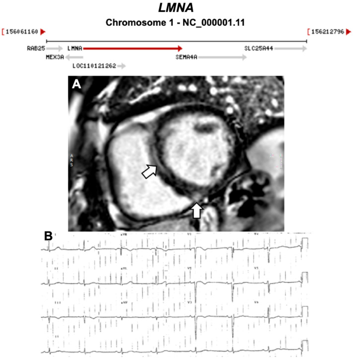
Figure 10.
Exemplar MRI and ECG finding found in ACM patients with LMNA mutations. (A) CMR of a patient with LMNA mutation showing a mid-wall (LGE) scar in the septum and inferior RV; (B) 12-lead ECG of a different patient with a p.Gly382Val LMNA mutation, which shows sinus bradycardia, poor R-wave progression, T-wave inversion in V1–V3 leads. (A) reproduced with permission from Augusto et al. (2019) [39]; and (B) reproduced with permission from Quarta et al. (2012) [41].

