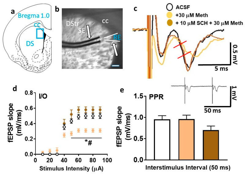
Figure 5.
Basal synaptic transmission is reduced by superfusion of methamphetamine in the dorsal striatum. (a) Schematic of a coronal slice representing the area of the dorsomedial striatum in blue rectangular box used for stimulation and recordings. (b) Infrared microphotograph of a 440-µm-thick corticostriatal slice from one adult male rat indicating the location of the stimulating electrode (SE) and recording electrode (RE). The scale bar in (b) is 100 µm. Thick arrows point to the electrodes. CC: corpus callosum; DStr: dorsal striatum. (c) A representative field excitatory postsynaptic potential (fEPSP) waveform illustrating the parameter computed in the study, including fEPSP slope (measured between the two red lines) from control- (black trace), methamphetamine- (beige trace), and SCH23390 + methamphetamine-treated (brown trace) slices. (d) Input/output (I/O) curve obtained by plotting the slope of fEPSPs as a function of the stimulation intensity (from 10 to 90 µA) in the dorsomedial striatum. (e) Paired-pulse ratio (PPR) from each slice is averaged from at least six traces with interstimulus interval at 50 ms. Inset shows an example trace used to calculate PPR. Data are represented as mean ± SEM. n = 9 control slices, n = 10 methamphetamine slices, and n = 5 SCH23390 + methamphetamine slices. * p < 0.05 vs. controls; # p < 0.05 vs. SCH23390 + methamphetamine by Fisher’s least significant difference (LSD) post hoc test.

