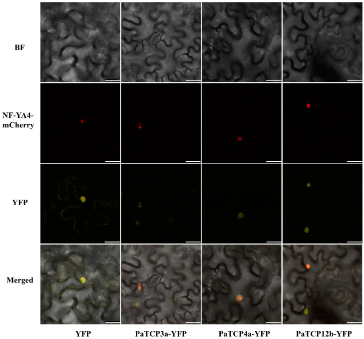
Figure 8.
Subcellular localization of three PaTCP-YFP fusion proteins in tobacco. Empty vector of yellow fluorescence (YFP) was used as control. The co-transformation of the nuclear marker (NF-YA4-mCherry) was used to visualize the nuclei. Merged panel showed the nuclear localization in tobacco epidermis cells. Scale bar, 25 μm.

