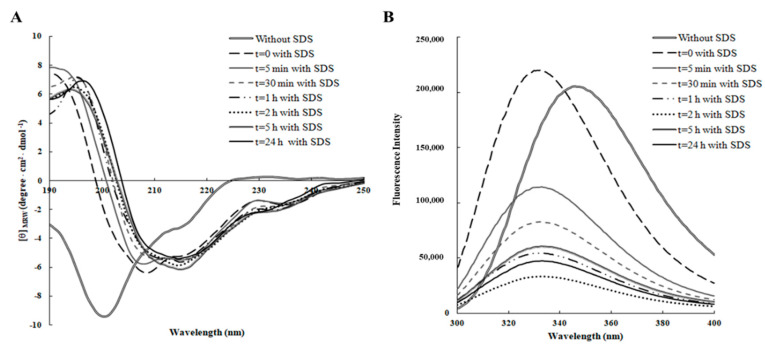
Figure 1.
Time-dependent effect of SDS on the secondary and tertiary structure of RiLK1 monitored by spectroscopic analyses. (A) Far-UV Circular Dichroism (CD) spectra of the peptide (0.1 mg/mL) were recorded in 10 mM Tris-HCl buffer pH 7.0 in the presence or absence of SDS (3 mM) over the time at 25 °C. (B) Intrinsic fluorescence emission spectra of RiLK1 (0.1 mg/mL) in 10 mM Tris-HCl buffer, pH 7.0 in the presence or absence of SDS (3 mM) over the time at 25 °C.

