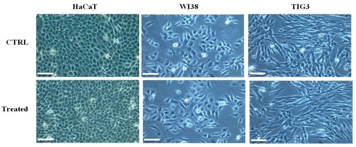
Figure 8.
Morphological observation of different human cell lines treated with RiLK1 under phase-contrast microscope. Keratinocyte (HaCAT), embryonic (WI38), and fetal (TIG3) lung fibroblastic cell lines were incubated at 37 °C for 24 h in absence (CTRL) or in presence (treated) of RiLK1 at the maximum concentration tested (10 µM). The microscope images are representative of three independent experiments performed in triplicate. Bar is equal 100 µm.

