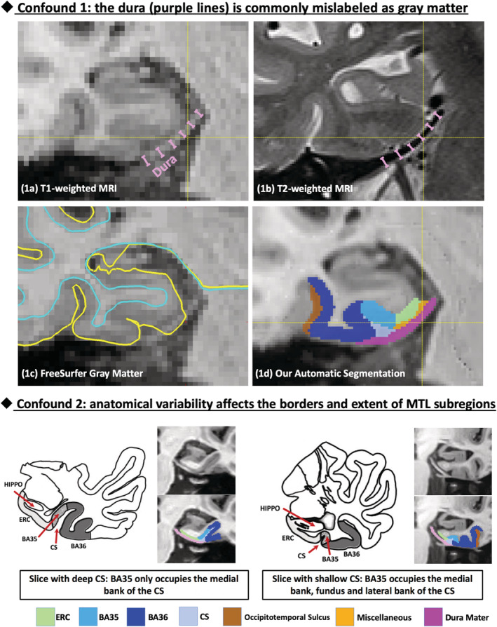
FIGURE 1.

Common confounds in automatic segmentation of medial temporal lobe (MTL) subregions using T1‐weighted MRI. Confound 1: the dura mater (indicated by purple lines) has similar intensity with gray matter (GM) in T1‐weighted MRI (a) but can be easily separated in T2‐weighted MRI (b), is commonly mislabeled as GM (c). Confound 2: Large anatomical variability exists in the MTL defined by the pattern of the collateral sulcus (CS), which influences the borders and extent of the subregions of the MTL cortex. Our segmentation pipeline is able to reliably separate dura from GM (1d) and account for anatomical variability. Figure adapted from Ding and Van Hoesen (2010). BA, Brodmann area; ERC, entorhinal cortex; HIPPO, hippocampus
