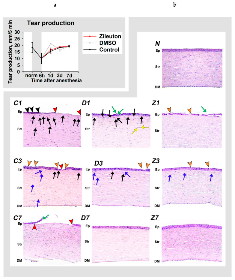
Figure 5.
Dynamic changes in tear secretion and corneal morphology in DES in the course of anti-inflammatory therapy. The animals were exposed to general anesthesia for 6 h (shown as gray box) and subsequently received instillations of the eye drops containing 50% DMSO (D1, D3, D7) or 50% DMSO with 0.5% zileuton (Z1, Z3, Z7) for 7 days. (a) The results of standardized Schirmer’s tests performed at 6th hour, 1st day, 3rd day or 7th day after the exposure. (b) Representative microscopic images of hematoxylin and eosin-stained cross-sections of the corneas at 1st (D1, Z1), 3rd (D3, Z3) and 7th (D7, Z7) day after exposure. The image of the normal cornea is also presented (N). Stromal denudation (complete absence of the corneal epithelium) areas (red arrowheads), loci of desquamation/destruction of the epithelial layer (orange arrowheads), and sites of reepithelialization (green arrows) are indicated. The inflammatory changes include granulocytic infiltration in the corneal epithelium (intraepithelial granulocytes; black arrowheads) and stroma (black arrows), neovascularization (yellow arrows), and postinflammatory signs of regeneration (activation of stromal keratocytes) at the ex-sites of the infiltration (blue arrows). Ep: epithelium; Str: stroma; DM: Descemet’s membrane and endothelium. Hematoxylin and eosin staining, magnification 200×. The central areas of the cornea are presented. Gaps, cracks and folds of preparations are of an artificial origin.

