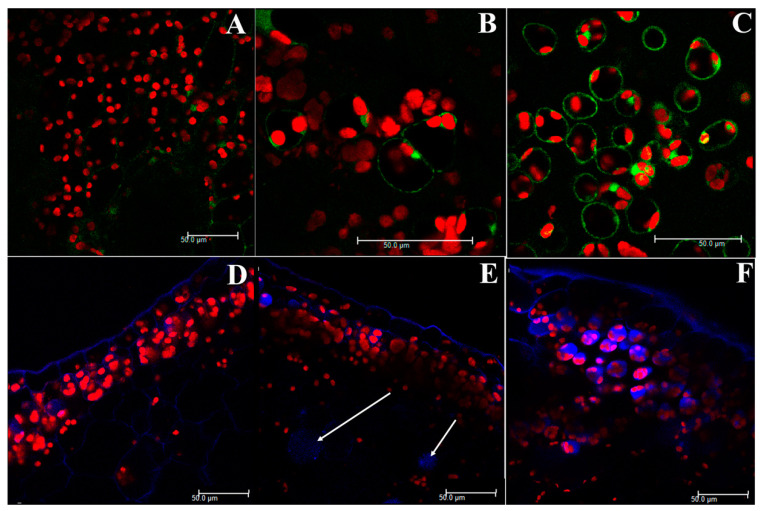
Figure 1.
Upper thalli of C. conicum samples observed under the confocal laser microscope, after dichlorofluorescein (DFC)-labelling (Figure 1A–C) and monochlorobimane (MCB)-labelling (Figure 1D–F). (A) C. conicum sample from the control culture, with a faint green light and a clear red light emission from chloroplasts. (B) C. conicum sample from the 36-µM Cd culture, with a visible green light emission from the cytoplasm. The red light from chloroplasts is comparable to control. (C) C. conicum sample from 360 µM Cd culture, with clear green and red light signals. Green light is collected from the cytoplasm, red light labels the chloroplasts. (D) C. conicum sample from control culture, with a blue light from the upper epidermis cell walls and a clear red signal from the chloroplasts. (E) C. conicum sample from the 36-µM Cd culture, with a blue light from the upper epidermis cell walls and a clear red signal from the chloroplasts. Cell vacuoles emit faint blue light (arrows). (F) C. conicum sample from the 360 µM Cd culture, with a strong blue signal from cell vacuoles, blue light from the upper epidermis cell walls and a clear red signal from the chloroplasts. Scale bars: 50 µm.

