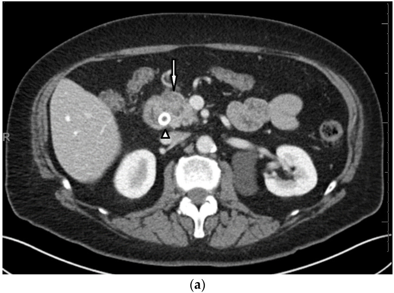
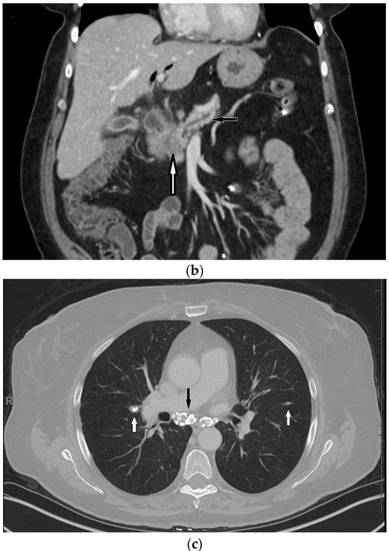
Figure 1.
Axial (a) and coronal (b) intravenous contrast-enhanced CT scans of the chest, abdomen, and pelvis at initial presentation show a 3.1 × 2.1 cm hypo-enhancing mass in the head of the pancreas (white arrows) with mild upstream pancreatic ductal dilatation and parenchymal atrophy (black arrow). The dilated biliary tree has been decompressed via a metal common bile duct stent (white arrow head); (c) Images through the chest on lung windows show calcified mediastinal lymph nodes (black arrow) and calcified pulmonary nodules (white arrows)—non-specific sequela of prior granulomatous process, including sarcoid.


