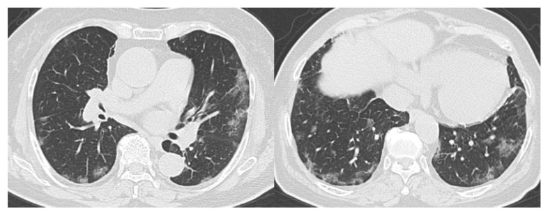
Figure 4.
Representative CT images of COVID-19 pneumonia (Case 2). Seventy-seven-year-old female. The CT images show multifocal bilateral, peripheral/subpleural ground-glass opacities with curvilinear bands, and subpleural sparing. RT-PCR was positive for SARS-CoV-2. All readers diagnosed it as Category 5. COVID-19, coronavirus disease 2019; CT, computed tomography; RT-PCR, reverse transcription-polymerase chain reaction; SARS-CoV-2, severe acute respiratory syndrome coronavirus 2.

