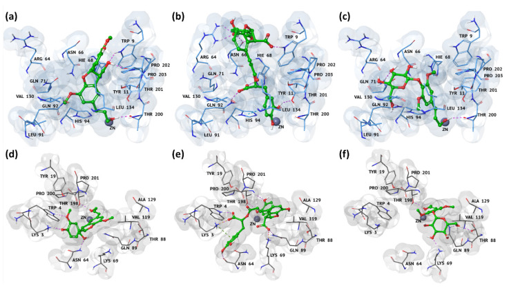
Figure 2.
Three-dimensional representations of the binding mode of (a–d) 6, (b–e) 12, and (c–f) 14’s best hits against hCA IX and XII binding pockets, respectively. The ligands are depicted as green carbon sticks, while hCA IX and XII are shown as blue and gray transparent cartoon, respectively. The zinc cation is represented as a violet sphere; the enzyme residues, involved in crucial contacts with the compounds, are reported as blue and gray carbon sticks, respectively, for hCA IX and XII isoforms. Hydrogen bonds, salt bridges, cations, and stacking interactions are reported, respectively, as dashed pink, orange, green, and light-blue lines. These binding modes derived from the molecular mechanics energy minimization performed by means of the eMBrAcE tool.

