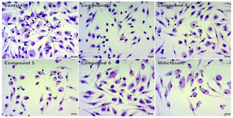
Figure 2.
Light microscopy of macrophages infected and treated with 1,4-disubstituted-1,2,3-triazole compounds 2, 4, or 5 at 37.5 μM; with compound 6 at 18.7 μM; or with miltefosine at 25 μM. Intracellular amastigotes (black arrows) and remains of amastigotes (red arrows) inside macrophages. Giemsa, 40× objective. The images are representative of two independent experiments performed in quadruplicate.

