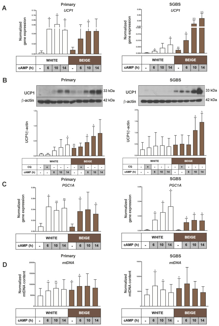
Figure 1.
The expressions of Uncoupling protein 1(UCP1), Peroxisome Proliferator-Activated Receptor Gamma Coactivator 1-Alpha (PGC1A) and mitochondrial DNA (mtDNA) were elevated upon cAMP stimulation. Abdominal subcutaneous hASCs and SGBS cells were differentiated to white or beige adipocytes for 2 weeks. A quantity of 500 µM dibutyryl-cAMP was administered for 6 to 14 h to induce thermogenesis, and chloroquine (CQ) treatment (25 µM, 1 h) was applied to block the lysosomal degradation activity. (A) UCP1 gene expression in primary (left) and Simpson–Golabi–Behmel syndrome (SGBS) cells (right) normalized to GAPDH, quantified by RT-qPCR (n = 5). (B) Representative immunoblots and densitometry analysis of UCP1 protein expression in hASC-derived (left) and SGBS cells (right) normalized to β-actin (n = 5). (C) PGC1A gene expression in primary (left) and SGBS cells (right) normalized to GAPDH quantified by RT-qPCR (n = 5). (D) Normalized mitochondrial DNA content quantified by qPCR in hASC-derived (left) and SGBS cells (right) (n = 6). Results are expressed as mean ± SD. Statistics: two-tailed paired student t-test; * p < 0.05; ** p < 0.01; * represents significant as compared to white untreated sample; △ p < 0.05; △△ p < 0.01; △ represents significant as compared to beige untreated sample.

