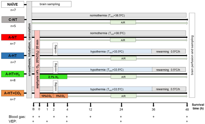
Figure 1.
Overview of the experimental protocol. Untreated animals served as naïve controls for neuropathology and gene expression studies; piglets in the control-normothermia group (C-NT) were anesthetized, ventilated, and monitored but not subjected to asphyxia. Animals of the asphyxia-normothermia group (A-NT) were exposed to 20 min asphyxia induced by ventilation with 4% O2–20% CO2 gas mixture. Animals in the asphyxia-hypothermia group (A-HT) were cooled down to 33.5 °C, starting 2 h after reventilation, and were gradually rewarmed from 36 h to normothermia by 0.5 °C/h in 10 h. H2-treatment was initiated at the onset of reventilation after asphyxia (21%O2–2.1% H2, 4 h) and was combined with hypothermia (A-HT + H2 group). In a similar fashion, CO2-treatment was initiated at the onset of reventilation after asphyxia and was combined with hypothermia (A-HT + CO2 group). In order to achieve graded restoration of normocapnia, the animals were ventilated first with 21% O2–10% CO2 for 2 h and then with 21% O2–5% CO2 for 2 h before switching back to air. Arterial blood samples were collected and visual evoked potential (VEP) measurements were performed at the marked time points.

