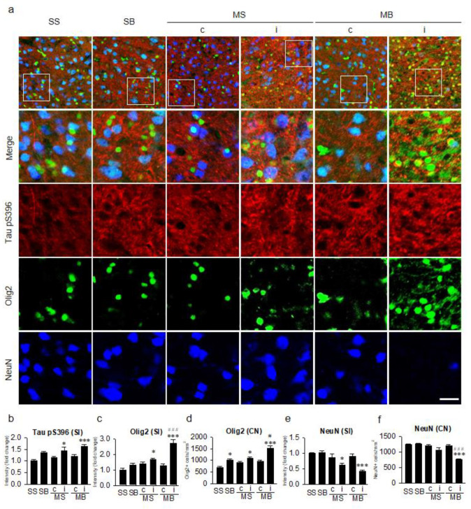
Figure 3.
Tauopathy in the cortex. (a) Triple-label immunohistochemistry of tau hyperphosphorylation at the serine 396 residue (Tau pS396) in conjunction with oligodendrocytes (Olig2) and neurons (NeuN). Quantitative analysis of the signal intensities of Tau pS396 (b), the signal intensities of Olig2 (c), the number of Olig2-positive cells (d), the signal intensities of NeuN (e), and the number of NeuN-positive cells (f). n = 6 per group; scale bar = 20 μm; SS, sham + sham; SB, sham + BCCAo; MS, MCAO + sham; MB, MCAO + BCCAo; MCAO, middle cerebral artery occlusion; BCCAo, bilateral common carotid artery occlusion; c, contralateral; i, ipsilateral; SI, signal intensity; CN, cell number; * p < 0.05 and *** p < 0.001 compared to SS; # p < 0.05 and ### p < 0.001 compared to the ipsilateral MS.

