Figure 2.
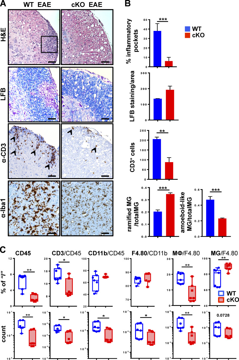
Histological signs of EAE are attenuated in spinal cords of GRIP1-cKO mice. (A) Spinal cord lumbar sections from WT and GRIP1-cKO mice at DPI20 from three independent experiments were analyzed by H&E staining for inflammatory foci (rectangle), LFB staining for myelin, and immunohistochemistry for CD3+ infiltrating T cells (arrowheads) and Iba-1+ MG (in parenchyma). Scale bars are 100 µm. (B) Quantification of slides from A for inflammation (percentage of inflammatory pockets), demyelination (LFB-stained myelin/area), CD3+ cells, and a number of ramified (with processes) and amoeboid-like (round-shaped with retracted processes) MG was performed as described in the Materials and methods section (unpaired two-tailed Student’s t test). **, P < 0.01; ***, P < 0.001. (C) FACS analysis of leukocytes isolated from spinal cords of WT or GRIP1-cKO mice at DPI20 is plotted as a percentage of gated parent population and total counts (Mann-Whitney U test). *, P < 0.05; **, P < 0.01.

