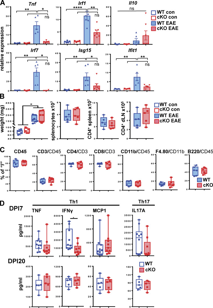
Figure 3.
GRIP1-cKO mice develop less brain inflammation than WT mice but a similar peripheral T cell response in vitro. (A) Brains were harvested from control (WT = 5; GRIP1-cKO = 4) and EAE DPI20 (WT = 6; GRIP1-cKO = 6) mice from two independent experiments, and total RNA was extracted. Relative expression of the indicated genes was evaluated by RT-qPCR, normalized to that of the Actb housekeeping gene, and expressed relative to WT control (= 1; two-way ANOVA with Tukey’s multiple comparisons test). *, P < 0.05; **, P < 0.01; ****, P < 0.0005. ns, nonsignificant. (B) Spleens were collected from WT and GRIP1-cKO control mice (n = 5 each) and EAE DPI20 mice (n = 7 each) from one experiment, and their weights (in mg) were compared (Kruskal-Wallis test with Dunn’s multiple comparisons test; *, P < 0.05). Numbers of splenocytes and CD4+ cells isolated from spleens and dLNs were quantified by FACS analysis. (C) FACS analysis of leukocytes isolated from spleens of WT and GRIP1-cKO mice at DPI20 is plotted as a percentage of the gated parent population. (D) Spleens were collected from WT and GRIP1-cKO mice at DPI7 (WT = 10; GRIP1-cKO = 9 from two independent experiments) and DPI20 (n = 5 each from one experiment). CD4+ T cells were isolated and restimulated with MOG35–55 in vitro, and the indicated Th1 and Th17 secreted cytokines were quantified using CBA (unpaired two-tailed Student’s t test). *, P < 0.05.

