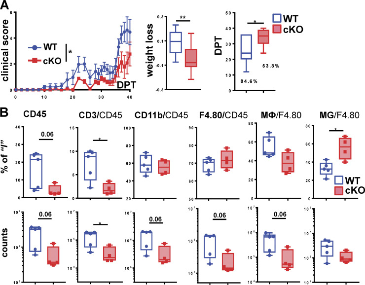
Figure 4.
GRIP1 contributes to the neuroinflammatory phase of EAE. (A) Passive EAE was induced in WT and GRIP1-cKO mice (n = 13 each from two independent experiments), and clinical scores (left) were measured and plotted daily as mean ± SEM (Mann-Whitney U test). The fraction of weight lost between days post-transfer (DPT) 0 and 40 (middle) was compared using an unpaired two-tailed Student’s t test. The incidence of disease (%) is shown; time of symptom onset (right) was compared using the Mann-Whitney test. *, P < 0.05; **, P < 0.01. (B) FACS analysis of leukocytes isolated from spinal cords of five WT and four GRIP1-cKO mice (two independent experiments) at DPT40 is plotted as a percentage of the gated parent population and total counts (unpaired two-tailed Student’s t test). *, P < 0.05.

