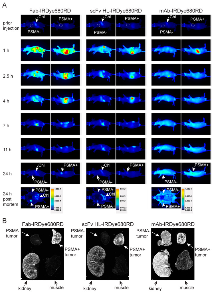
Figure 5.
Pharmacokinetics of 5D3 variants and ex vivo NIRF imaging. (A) Mice bearing a PSMA-positive (PSMA+, right side) and PSMA-negative (PSMA−, left side) xenograft (location of tumor marked by dashed circle), were intravenously injected with 5D3 variants conjugated to IRDye680RD and images collected at various times post-injection with identical exposure settings. Both Fab and scFv fragments revealed specific localization to the PSMA-positive tumor already after 2.5–4 h p.i., while non-specific binding was still observed for 5D3 mAb at 24 h p.i. due to high levels of the circulating conjugate. At 24 h p.i. mice were sacrificed, and the ventral part dissected, and mice were scanned. The fluorescent signal in the gastrointestinal tract originates from chlorophyll (Chl) present in feed. Figures are representative images from triplicate runs for each 5D3 variant. (B) Sections of tumors, kidney and skeletal muscle (24 h p.i.) were scanned ex vivo. The specific signal for scFv and Fab was localized mainly to the rim of PSMA-positive tumors and to the kidney whereas PSMA-negative tumors and skeletal muscle did not show any significant signal. 5D3 mAb showed a strong signal in both tumors as well as kidney, and also observable signal in the PSMA-negative tumor and skeletal muscle.

