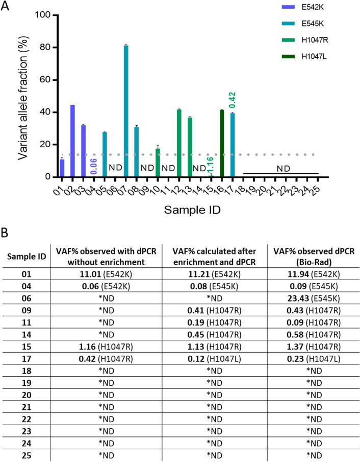
Figure 4.
PIK3CA mutation detection in breast cancer FFPE biopsy tissue. (A) Genomic DNA isolated from FFPE tissue biopsy samples from a total of 25 breast cancer patients were subjected to dPCR analysis for PIK3CA E542K, E545K, H1047R and H1047L mutations. Data are shown as VAF for a detected target mutation. All samples with VAF < 15% (below the dotted line) were then enriched prior to dPCR analysis. Sample 17 was included in the exon 20 enrichment experiment due to low VAF of H1047R. The results are presented with their Poisson-based 95% confidence intervals. (B) The logarithmic regression method was applied to calculate initial VAF for FFPE tissue biopsy samples after enrichment with nuclease-based assays and detection with dPCR. Calculated initial VAF% results were compared to dPCR prior to enrichment and ddPCR results obtained at Institute of Cancer Research, London, UK, using a commercial Bio-Rad system and larger quantities of sample template DNA. *ND—not detected.

