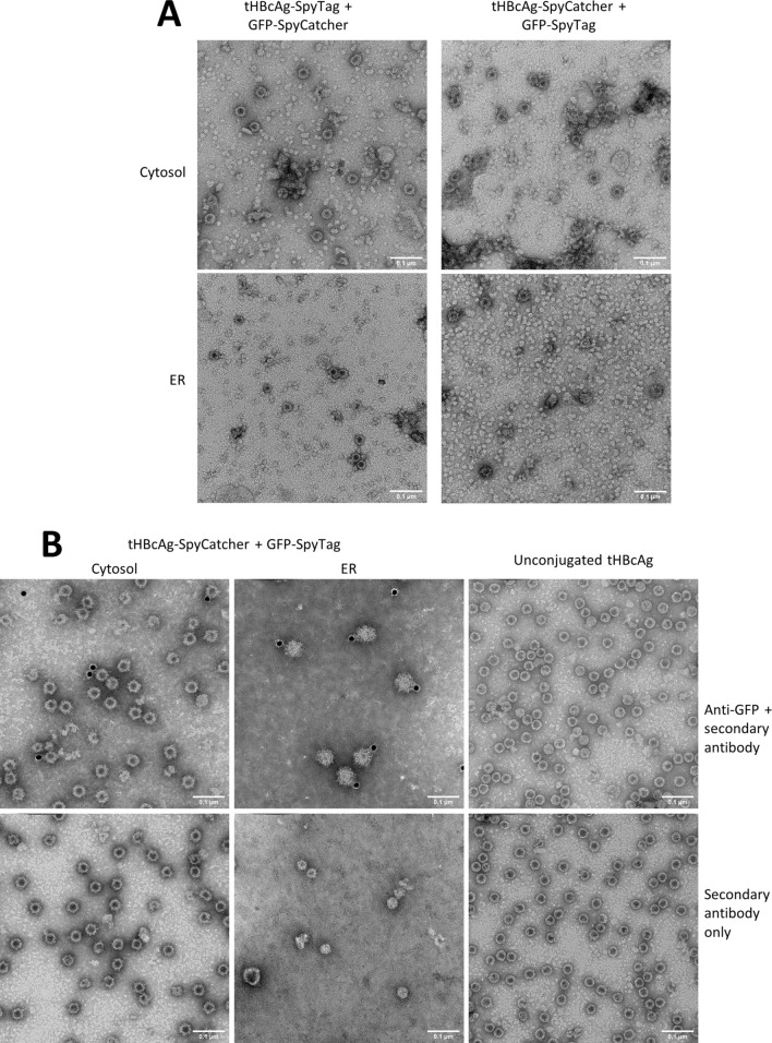
Figure 6.
Transmission electron microscopy analysis of plant-produced VLPs. (A) 70% sucrose fractions. All constructs led to the assembly of tHBcAg VLPs in a comparable size and shape as previously published28. (B) Immunogold labelling of Nycodenz gradient-purified VLPs. Top row: grids incubated with anti-GFP primary antibody then gold-conjugated secondary antibody. Bottom row: grids incubated with gold-conjugated secondary antibody only. All samples were stained with 2% (w/v) uranyl acetate, scale bars are 100 nm each.

