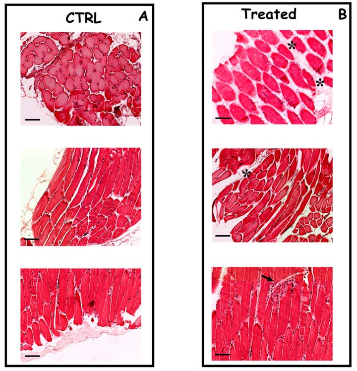
Figure 3.
Histopathological analysis of skeletal muscle of mice untreated or treated by needle electrodes. Panel (A) cross-sections of skeletal muscle from an untreated mouse, showing no significant histopathological alterations (hematoxylin and eosin, original magnification ×20); Panel (B) cross-sections of skeletal muscle from a mouse treated by caliper electrodes showing a focus of mild mononuclear inflammatory infiltrate (arrow) and a slight deviation of the fibers (asterisks). (hematoxylin and eosin, original magnification ×20). Scale bar represents 100 μm for all images.

