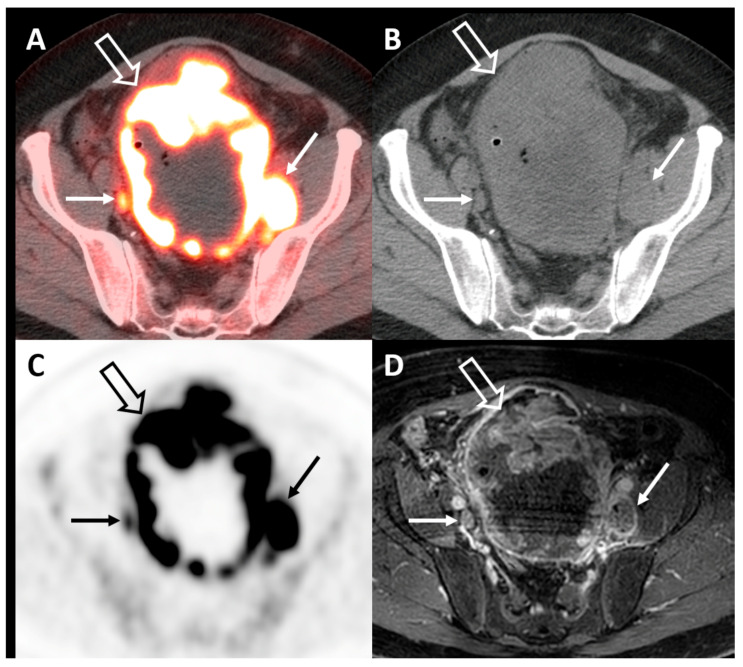
Figure 3.
Axial fused FDG-PET/CT (A), CT (B), PET (C), and corresponding T1-fat saturated postcontrast MRI (D) demonstrating an extensive hypermetabolic mass involving nearly all of the bladder wall (open arrow) and multiple pelvic lymph node metastases (closed arrow), some of which measure less than 1 cm in short axis and are more easily identified on PET/CT vs. CT or MRI alone.

