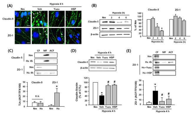
Figure 3.
Effects of yuzu and HSP on disruption of claudin-5 and ZO-1 during hypoxia in bEnd.3 cells. (A) Representative fluorescence images of disruption of claudin-5 and ZO-1 induced by 4 h of hypoxia in brain endothelial cells. Arrows indicate regions of tight junction protein disruption. Green: claudin-5- or ZO-1-conjugated FITC, blue: Hoechst. Scale bar, 20 µm. (B–E) Western blotting of claudin-5 and ZO-1. (B) Protein levels of claudin-5 and ZO-1 during hypoxia (2, 4, and 6 h). (C) Redistribution of claudin-5 and ZO-1 during at 4 h of hypoxia in each fraction of bEnd.3 cells. (D) Effect of yuzu and HSP on degradation of claudin-5 at 4 h of hypoxia. (E) Effect of yuzu and HSP on redistribution of ZO-1 at 4 h of hypoxia. Data are shown as mean ± SEM. (n = 5–7) * p < 0.05 vs. Nm, # p < 0.05 vs. Veh. Nm: normoxia; Veh: vehicle; H: hypoxia; HSP: hesperidin; CF: cytosolic fraction; MF: membrane fraction; ACF: actin cytoskeleton fraction.

