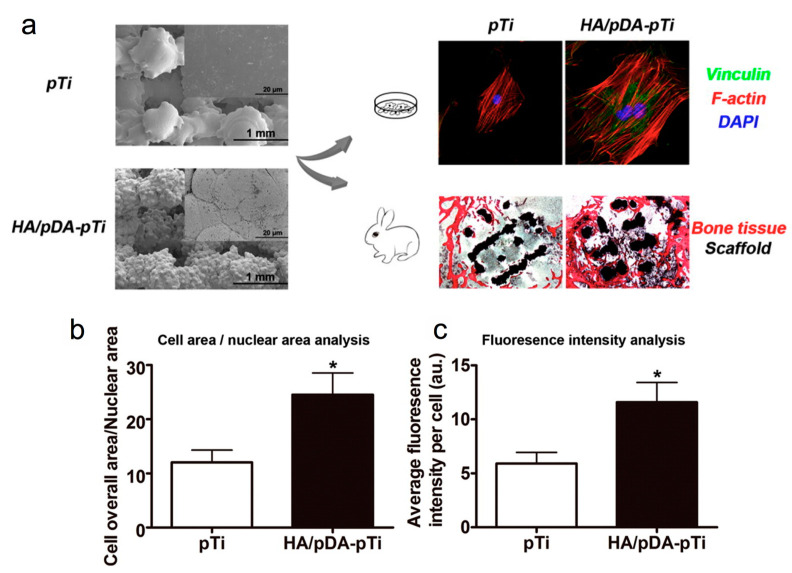
Figure 5.
(a) Scanning electron microscopic (SEM) images of bare porous Ti6Al4V scaffolds (pTi) and polydopamine-assisted hydroxyapatite coating on titanium surfaces (HA/pDA-pTi). Fluorescent staining of MC3T3-E1 cells adhered to pTi and HA/pDA-pTi scaffolds, and (b) analysis of morphology of SEM images as shown in panel a and (c) fluorescence intensity of vinculin staining for cells on different scaffolds. Asterisks (*) indicate statistical significance compared to the pTi group, p < 0.05). Reprinted with permission from reference [55]. Copyright (2015) American Chemical Society.

