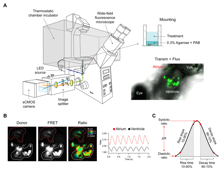
Figure 1.
Zebrafish embryo mounting for microscopy, image acquisition and processing, and parameters extracted from Ca2+ transients. (A) Embryos expressing the biosensors in the heart were embedded in agarose and mounted in glass-bottom 96-well plates. An overlay of transmitted light and fluorescence of the heart is shown in an embryo expressing Twitch-4. The fluorescence from the widefield microscope passed through an image splitter that separated the FRET and donor emission onto the sCMOS sensor. (B) The two emission images were divided off-line and the ratio images (FRET/donor channels) were computed. Regions-of-interest (ROI) were manually drawn on the atrium (red line) and ventricle (white line) and analyzed in Igor Pro (WaveMetrics). (C) Various parameters were automatically extracted from the ratio time course data.

