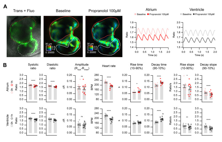
Figure 7.
In vivo Ca2+ kinetics in the heart of Twitch-4-expressing embryos in response to propranolol. Imaging was performed before and after 1 h incubation with 100 µM propranolol in 3 dpf zebrafish embryos expressing Twitch-4 in the heart. (A) Overlay of transmitted light and fluorescence in a representative experiment (left image). The corresponding emission ratio images are shown in pseudo color before (middle) and after (right) 1 h incubation with propranolol. ROIs were drawn to delimit fluorescent cells in the atrium (red) and ventricle (white). The graphs show the oscillations of Ca2+ in the atrium and ventricle before and after propranolol. (B) Effect of propranolol on the kinetic parameters of the cardiac Ca2+ transients. Data are shown as the mean ± S.E.M., n = 11 and 13 embryos for atrium and ventricle, respectively, from two independent experiments. A paired Student’s t-test was used (* p < 0.05, ** p < 0.01, **** p < 0.0001, n.s. not significant).

