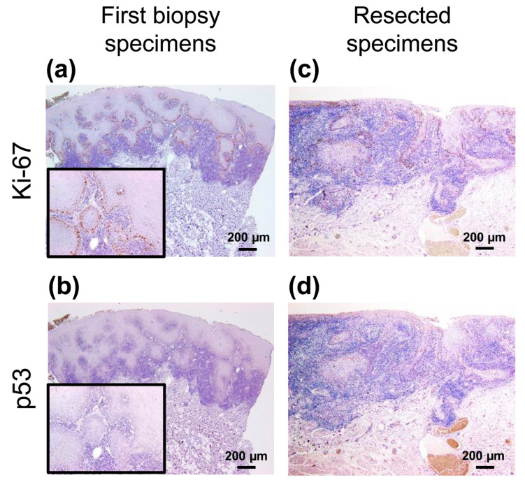
Figure 2.
Immunohistochemical analyses of the first biopsy and resected specimens. (a,c) Ki-67+ cells were mainly observed in the basal cell layer in both specimens. (b,d) Few p53+ cells were confirmed in both the specimens. Scale bars in (a)–(d) = 200 µm. Insets in (a) and (b) show high power view of each images.

