Figure 5.
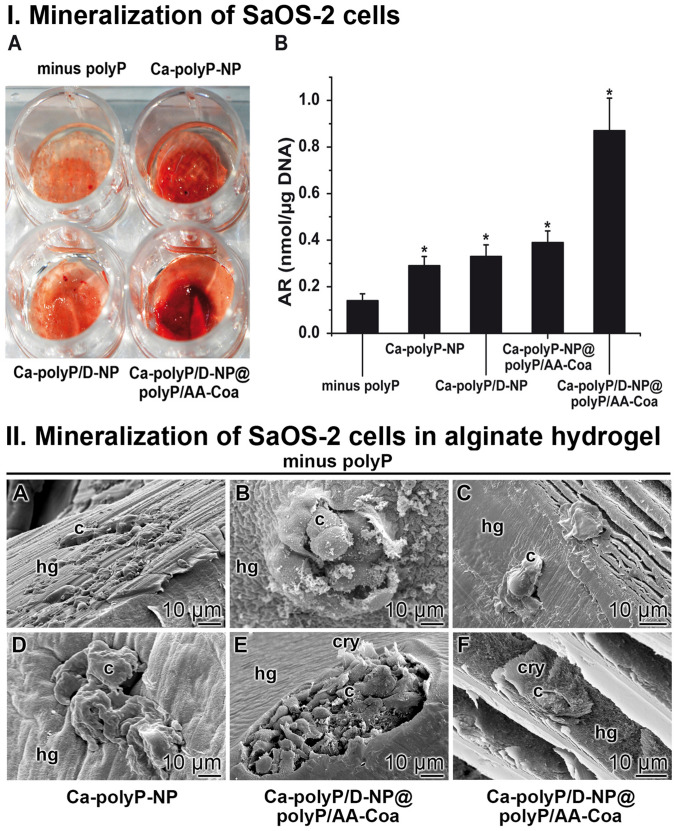
Effect on cell surface mineralization. I. Mineralization of SaOS-2 cells within an alginate hydrogel supplemented with different formulations of polyP. (I-A) The cells were embedded into the alginate either in the absence of polyP (minus polyP) or into a matrix containing either “Ca–polyP-NP” or the DEX-enriched particles, “Ca–polyP/D-NP”, as well as “Ca–polyP/D-NP@polyP/AA-Coa”. At the end of incubation (5 d) the gel was colored with Alizarin Red S. (I-B) Quantitative assessment of mineralization onto SaOS-2 cells, embedded into the hydrogel, either in the absence of the polymer (minus polyP), or in the presence of the polymer in the form of “Ca–polyP-NP”, “Ca–polyP/D-NP”, “Ca–polyP-NP@polyP/AA-Coa” or “Ca–polyP/D-NP@polyP/AA-Coa”, as described under “Materials and methods”. The determinations were performed after 5 days with Alizarin Red S. The signals determined were normalized to allow correlation with the cell numbers. Means ± SD; n = 10; *, p < 0.005). II. Visualization of SaOS-2 cells within the alginate-based hydrogel. The cells were cultivated within the hydrogel (A to C) in the absence of polyP, (D) in “Ca–polyP-NP”-enriched gel, or (E and F) in “Ca–polyP/D-NP@polyP/AA-Coa”-enriched gel; ESEM. The cells were seeded into the respective hydrogel (hg) and incubated for 5 days. The cells (c) and crystallites (cry) on their surfaces are marked.

