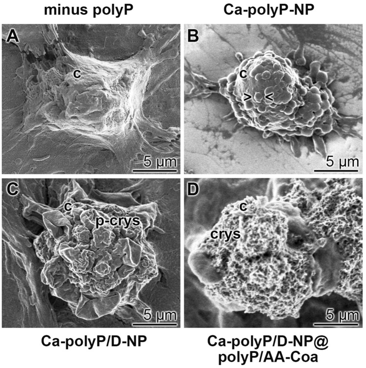
Figure 6.
Change of the morphology of the SaOS-2 cell surface in dependence on the incubation condition within the alginate hydrogel after an incubation for 5 days. Representative images are shown; SEM. (A) In the absence of polyP in the gel the cell surface is smooth. This morphology alters after exposure of the cells to the gel containing polyP. (B) In gel containing “Ca–polyP-NP” the cell surface undulates > < ; (C) after exposure to “Ca–polyP/D-NP” the cells start to form pre-crystallites (p-crys) and (D) after incubation with “Ca–polyP/D-NP@polyP/AA-Coa” the surface is studded with crystallites (crys).

