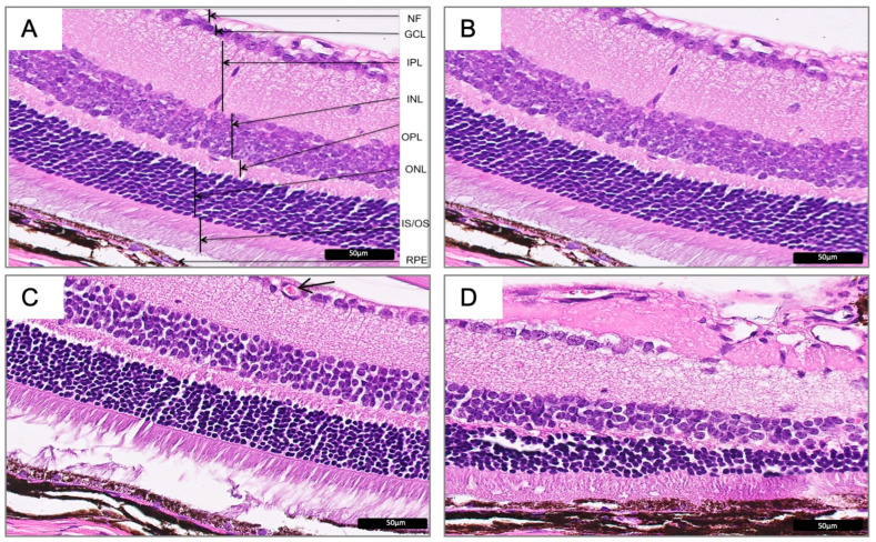
Figure 2.
Hematoxylin and eosin images representing normal mice retina in Wfs1-wild-type mice (A,B) and the corresponding representative images of the defect mice retina in the Wfs1-mutated mice (C,D). The neovascularization (C, arrow) and morphological (D) defects can be seen. Magnification of 400X. Abbreviations: Wfs1: Wolfram syndrome 1 gene mice; NF: nerve fiber layer; GCL: ganglion cell layer; IPL: inner plexiform layer; INL: inner nuclear layer; OPL: outer plexiform layer; ONL: outer nuclear layer-representing photoreceptor cell bodies; IS/OS: inner segment/outer segment; RPE: retinal pigment epithelium.

