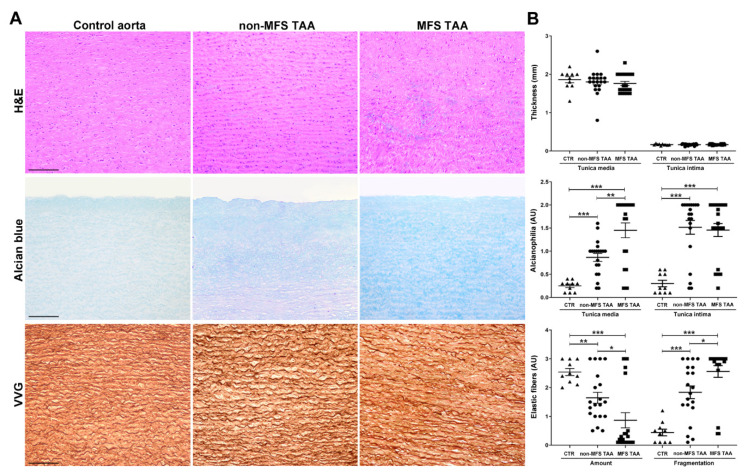
Figure 1.
Increased alcianophilia and elastic tissue remodeling characterize Marfan syndrome (MFS) thoracic aortic aneurysm. (A) Representative aortic media sections stained with hematoxylin and eosin (H&E) show greater degeneration with accumulation of basophilic material in the tunica media of MFS thoracic aorta aneurysm (TAA). Alcian blue staining shows increased accumulation of alcianophilic material in MFS compared to non-MFS TAA and control aorta. Verhoeff–Van Gieson (VVG) staining documents greater loss and fragmentation of elastic fibers and a more irregular arrangement in the tunica media of MFS compared with non-MFS TAA and control aorta. Scale bars equal to 150 μm. (B) Bar graphs of morphometric evaluations concerning intimal and medial thickness, alcianophilia and elastic tissue loss and fragmentation in non-MFS and MFS TAA and control aortas. MFS TAA (n = 20), non-MFS TAA (n = 20) and control aorta (n = 10). Average are reported as means ± SEM; * p < 0.05; ** p < 0.01; *** p < 0.001; estimated by t-test. Abbreviations: AU, arbitrary units.

