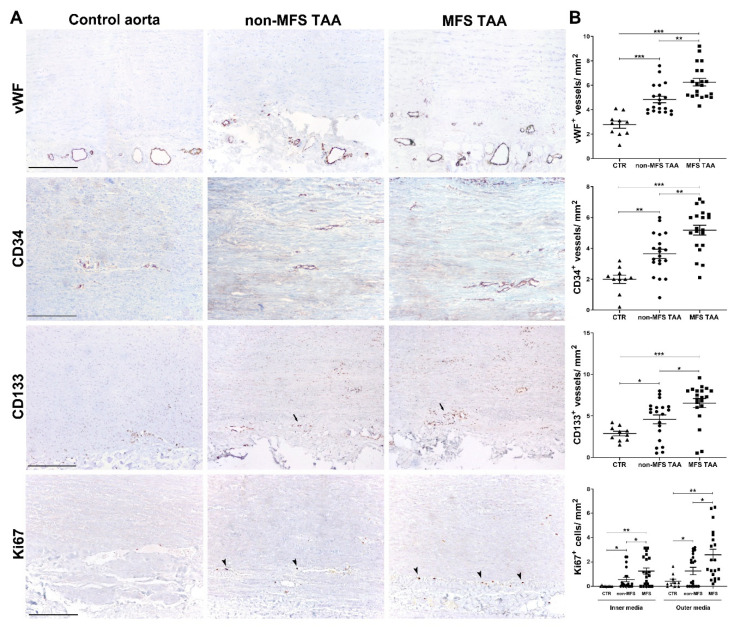
Figure 3.
Increased angiogenesis and vascular precursor cell recruitment characterize MFS thoracic aortic aneurysm. (A) Representative images of vWF, CD34, CD133 and Ki67 immunostainings in the tunica media of MFS and non-MFS TAA and control aorta. Perivascular CD133+ cells are indicated by arrows, whereas Ki67+ nuclei are represented by arrowheads. Scale bars are equal to 150 μm. (B) Semiquantitative evaluations of immunoreactivity show increased vWF, CD34+ and CD133+ neovessels, as well as a higher number of Ki67+ cells/ mm2 mostly in the outer media of MFS TAA compared with non-MFS and control aorta. MFS TAA (n = 20), non-MFS TAA (n = 20) and control aorta (n = 10). Averages are reported as means ± SEM; * p < 0.05; ** p < 0.01; *** p < 0.001; estimated by t-test. Abbreviations: AU, arbitrary units.

