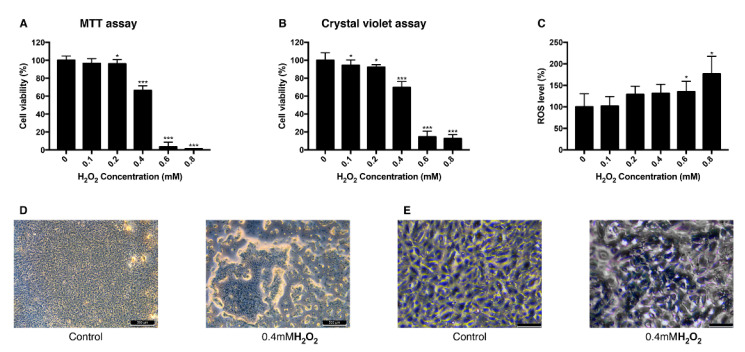
Figure 1.
Change of viability and intracellular ROS level in ARPE-19 cells after exposure to H2O2. The response of ARPE-19 cells to 0–0.8 mM H2O2 exposure for MTT assay (A), and crystal violet assay (B) to determine cell viability. For intracellular ROS level, DCFH-DA was treated for 30 min after the H2O2 exposure. Exposure to H2O2 reduced the cell viability (A,B) and increased the intracellular ROS level (C). The cell morphology was observed with bright field microscopy (Scale bar 500 μm) (D) and with higher magnification (scale bar 100 μm) (E). Asterisks indicate a significant reduction in cell viability or increment in ROS level compared with untreated cells (* p < 0.05, ** p < 0.01, *** p < 0.001). MTT, 3-(4,5-dimethylthiazol-2-yl)-2,5-diphenyltetrazolium bromide; ROS, reactive oxygen species; DCFH-DA, 2′,7′-dichlorodihydrofluorescein diacetate.

