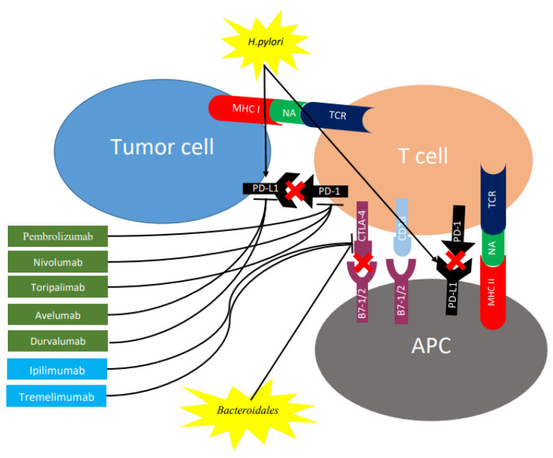
Figure 2.
Role of checkpoint inhibitors and gut microbiome on expression of CTLA-4 and PD-1 in regulating different stages of T cell response. T cell activation requires two complementary signals: The interaction between the TCR and peptide-MHC complex must be associated with a second co-stimulatory signal mediated by CD28. Conversely, the binding of CTLA4 to B7-1/2 provides a control signal that suppresses ongoing T cell activation. PD-1 is upregulated on T cells following persistent antigen exposure. When PD1 binds to its ligand, PD-L1 or PD-L2, expressed by tumor cells, the T cell receives an inhibitory signal. Antibodies against CTLA-4 (shown in blue rectangles) or PD-1/PD-L1 (shown in green rectangles) can activate T cells. H. pylori increases gastric epithelial expression of PD-L1 while bacteroides block CTLA-4 expression. CTLA-4, cytotoxic T lymphocyte antigen 4; PD-1, programmed cell death protein 1; TCR, T cell receptor; MHC, major histocompatibility complex; PD-L1, programmed death ligand 1; APC, antigen-presenting cell; NA, neoantigen.

