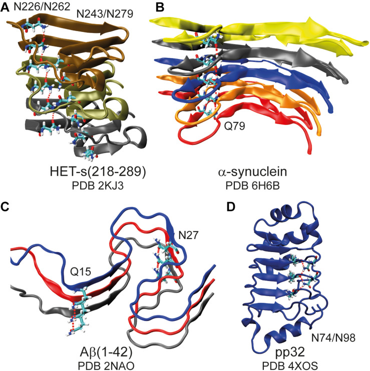
FIGURE 1.
Asparagine and glutamine ladders in protein structures. Schematic representation of amyloid fibrils (A–C) highlighting asparagine and glutamine ladders and a leucine-rich repeat protein in which two asparagine side-chains constitute to a second backbone (D). Hydrogen bonds are highlighted by red dashed lines. Note that for (A) structure 16 of the lowest energy structural bundle is shown as it particularly nicely illustrates the ladder motif (for more structures see Supplementary Figure S1).

