
Figure 6.
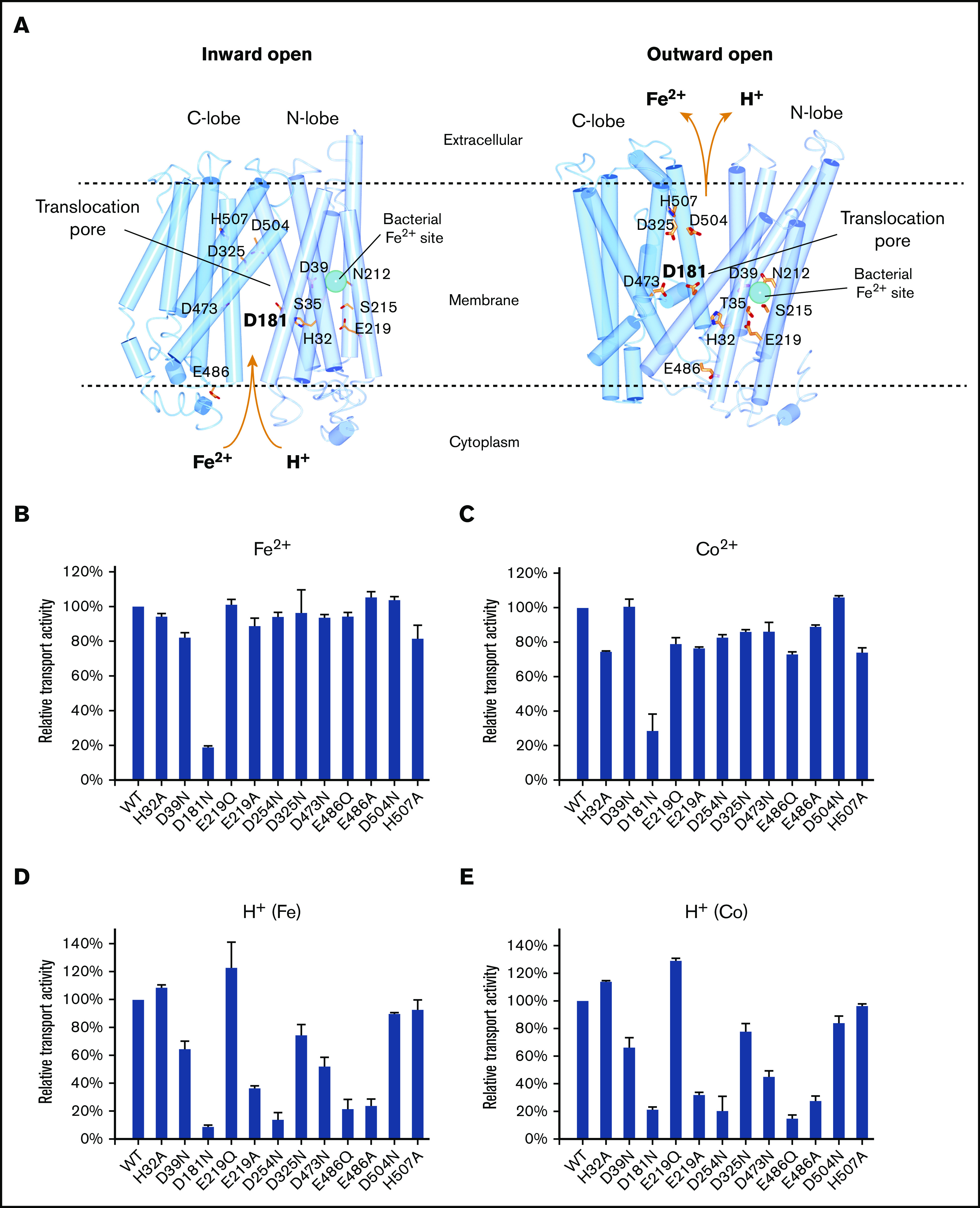
Iron and proton transport activities of ferroportin mutants. (A) Homology model of human ferroportin in inward-open (left) and outward-open (right) conformations. The models are generated by I-TASSER45 using the PDB code 5AYN and 5AYO. The iron-binding site in the bacterial homolog is shown in cyan sphere. (B-C) Relative Fe2+ and Co2+ transport activities of mutants. Experimental setups are the same as in Figure 1. (D-E) Relative proton transport activities of the mutants under a Fe2+ and Co2+ gradient. Experimental setups are the same as in Figure 4.